 EN
EN
 EN
EN AR
AR BG
BG HR
HR CS
CS DA
DA NL
NL FI
FI FR
FR DE
DE EL
EL HI
HI IT
IT JA
JA KO
KO NO
NO PL
PL PT
PT RO
RO RU
RU ES
ES SV
SV IW
IW ID
ID LV
LV LT
LT SR
SR SK
SK SL
SL UK
UK VI
VI ET
ET HU
HU TH
TH TR
TR FA
FA MS
MS BE
BE HY
HY AZ
AZ KA
KA MN
MN MY
MY KK
KK TG
TG UZ
UZ KY
KY
Comprehensive Guide to 6 Types of Acute Wounds, Pressure Ranges, Dressing Selection, and Clinical Pain Points
I. Why does the management of acute wound parameters require "differentiation-based treatment"?
Although acute wounds also fall under the category of newly developed wounds, there are significant variations in terms of exudate volume, depth of tissue injury, pain sensitivity, and the requirements for skin graft survival; thus, the use of uniform parameter settings represents one of the most common misconceptions in clinical practice. The expert consensus classifies recommendations into three levels—A, B, and C—based on the level of evidence: A = RCT (high-quality randomized controlled trial); B = cohort study; C = case series report. Gibson DJ's 2022 study demonstrated through biomechanical analysis that PU and PVA foams exhibit significant differences in liquid clearance rates and negative pressure transfer uniformity, and therefore should not be substituted arbitrarily.
Core Evidence-Based Rationale
Pressure gradient criteria: superficial wounds – 40––60 mmHg; deep wounds – 70––120 mmHg; the intermittent mode is recommended (negative pressure for 3–5 min, followed by a 2-min interval).
The drainage rate and negative pressure uniformity of PU foam are significantly superior to those of PVA; in conventional PVA foam, pore collapse occurs when the negative pressure exceeds 140 mmHg, and at 200 mmHg, 93% of samples exhibit uneven edge pressure distribution.
II. PU Black Foam vs. PVA White Foam: Dressing Selection Comparison Table
| Comparison Dimensions | PU Black Foam | PVA white foam |
| Liquid drainage capacity | Features the highest flow rate and is suitable for a wide range of exudative wounds. | The air diversion rate is relatively low, and high negative pressure may lead to collapse. |
| Negative pressure distribution profile | Uniform pressure distribution across the entire wound surface with no attenuation. | Negative pressure loss at wound edges was observed, with a significant difference exceeding 140 mmHg. |
| Wound adhesion properties | Granulation tissue tends to grow into the dressing, making wound dressing changes potentially painful and associated with bleeding. | Low-adhesive material that protects newly formed tissue and minimizes pain sensation. |
| Core Use Cases | Deep wounds, wounds with excessive exudate, or wounds requiring rapid granulation tissue formation | Superficial wounds, skin grafting sites, and pain-sensitive wounds |
| Recommended negative pressure range | -125~-175 mmHg | -125 to-175 mmHg; long-term values exceeding 140 mmHg are not recommended. |
Techniques for combining different dressings
For wounds with significant exudation and patients who are highly sensitive to pain: the inner layer consists of PVA white foam in direct contact with the wound surface, while the outer layer is wrapped with PU black foam, enabling both painless dressing changes and efficient drainage.
III. Quick Reference Guide to 6 Categories of Acute Wound Parameters & Key Clinical Implementation Points
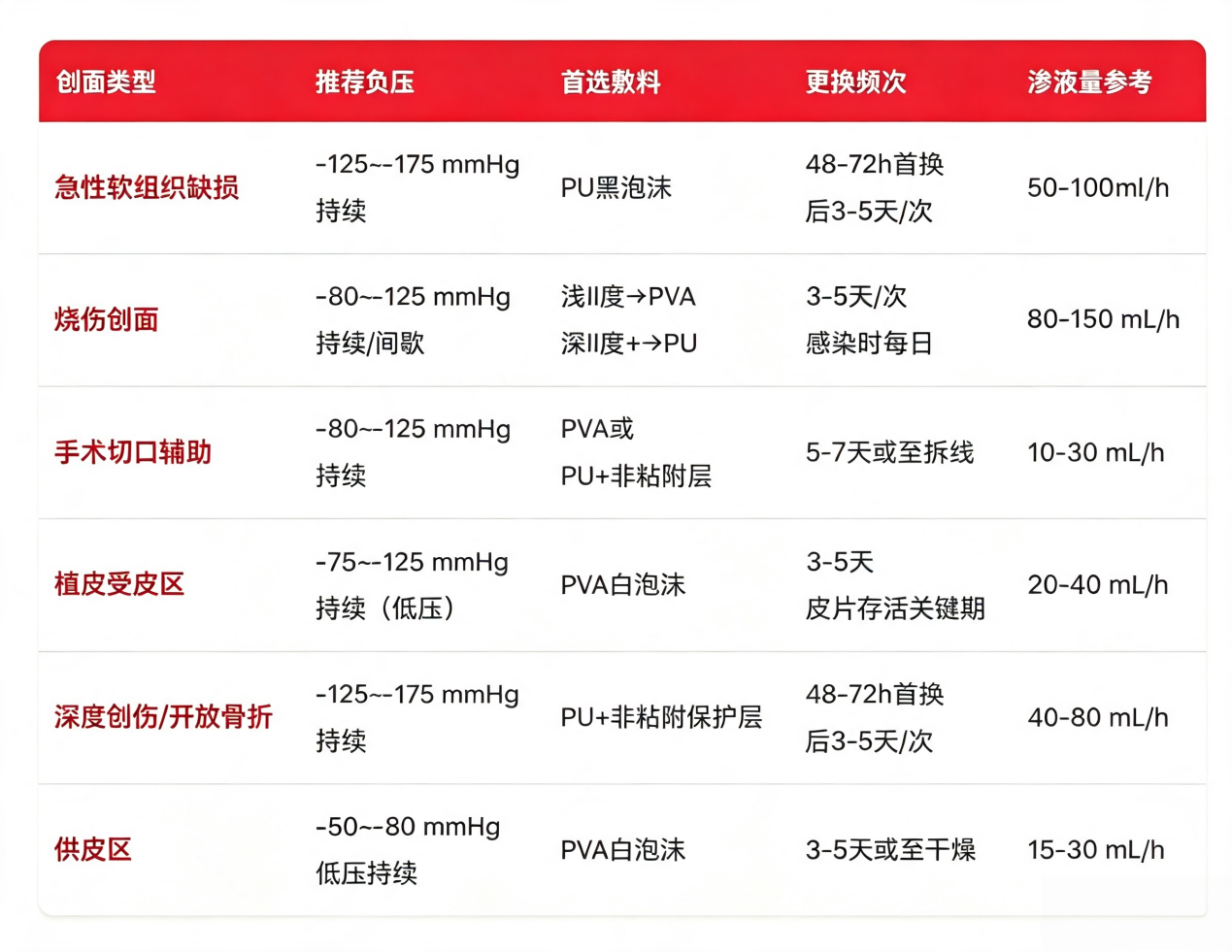
01 Traumatic skin/soft tissue defects (e.g., motor vehicle accidents, crush injuries, machinery entanglement injuries)
Negative pressure parameters: -125––175 mmHg, continuous negative pressure mode
Suture dressing selection: The preferred choice is PU black foam; for patients with pain sensitivity, the PVA inner layer combined with a PU outer layer configuration is recommended.
Clinical challenges: deep wounds with significant exudation; PU foam may promote granulation tissue growth, potentially leading to bleeding during dressing changes.
Evidence-based recommendations from expert consensus: deep wounds are recommended to use an intermittent mode with a pressure range of 70–120 mmHg; the standard reference negative pressure for the 3M device is –125 mmHg.
02 Burn wounds (II degree or higher burns)
Negative pressure range: Superficial II degree – 60––80 mmHg; Deep II degree/III degree – 80––125 mmHg; intermittent mode (negative pressure for 2–3 min, followed by a 1-min interval).
Dressing Layering Protocol: Superficial second-degree burns → PVA white foam, which can significantly reduce severe pain during dressing changes; deep second-degree/third-degree wounds after eschar removal → PU black foam, which enhances drainage and accelerates granulation tissue growth.
Wound care reminder: If signs of infection appear at the wound site, dressings must be changed daily; do not follow the conventional 3–5-day change interval.
03 Surgical incision (prophylactic NPWT for high-risk incisions)
Indications: Closed incisions for major orthopedic surgeries, abdominal surgeries in obese patients, and other procedures where seroma formation or infection is likely to occur.
Negative pressure parameters: conventional intervention – 50––70 mmHg; the standardized preventive system employs a sustained negative pressure of –125 mmHg.
Prioritize PVA white foam, as it will not damage the newly formed epidermis during removal; if PU foam is selected, a non-adhesive barrier layer must be added.
Recommended treatment course: Continue use for 5–7 days or until the sutures are removed from the incision; frequent dressing changes are not required.
04 Skin graft recipient site (thin-layer / full-thickness free skin graft)
Dynamic pressure-regulating protocol (maintaining sustained low pressure throughout the entire process): Postoperatively days 0–2: -25 to-75 mmHg; low-pressure protection is applied to the skin to prevent displacement; postoperatively days 2–12: -50 to-75 mmHg; the skin graft remains stable and adherent; postoperatively days 13–18: the pressure may be increased to-75 to-100 mmHg to promote basal healing.
Suture material selection: PVA white foam is the preferred choice; PU foam should be used with caution, as it can readily adhere to and tear the skin graft, leading to transplant failure.
Core risks: subcutaneous fluid accumulation, skin graft necrosis, and skin graft detachment resulting from improper dressing change procedures.
05 Open fracture / Deep complex trauma (with exposed bone or tendon)
Negative pressure parameters: -125––175 mmHg, continuous negative pressure mode; suitable for hemodynamic exudate at flow rates of 40–80 mL/h.
Choosing Dressings:PU black foam + silicone/olefin barrier protective layer; the barrier layer isolates exposed bone or tendon areas, preventing tissue ingrowth into the dressing.
Advanced treatment regimen: For large bone defect wounds, a combination of artificial dermal matrix and NPWT can be employed to promote granulation tissue coverage.
06 Donor site wound
Negative pressure parameters: The clinically preferred setting is the low-pressure continuous mode – 50––80 mmHg, which provides maximal relief of severe pain; according to expert consensus, the recommended baseline range for the intermittent mode is –70––120 mmHg.
Wound dressing selection: The only recommended option is PVA white foam, which prevents secondary injury caused by dressing adhesion.
Wound characteristics: moderate exudation (15–30 mL/h); severe pain is the patient's primary complaint.
IV. Three-Statement Clinical Quick Summary
Rule of thumb for dressing selection: For high fluid loss, choose PU; for wounds that are sensitive to pain, choose PVA; when both requirements need to be met, these two types can be used in combination through layered application.
Negative pressure threshold: For PVA foam dressings, prolonged application of negative pressure exceeding 140 mmHg is not recommended; for superficial wounds, skin grafting sites, and donor sites, low negative pressure is preferred; for deep wounds, the negative pressure may be moderately increased.
Standard treatment protocol: First, assess the depth of the wound and the amount of exudate; next, select the appropriate dressing; finally, apply negative pressure—ensuring that no single set of parameters is applied uniformly to all wounds.
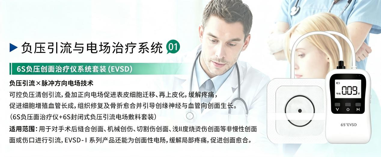
V. Product Introduction: Hairun Biotech's 6S Negative Pressure Wound Therapy System (EVSD)
Traditional negative pressure drainage can only remove exudate and reduce edema; in contrast, the Hairun 6S device innovatively combines negative pressure drainage with a pulsed DC electric field dual-technology, leveraging this dual mechanism to accelerate wound healing:
The functions of negative pressure technology include: eliminating wound edema, removing lactate and exudates from the wound surface, creating a moist healing microenvironment, as well as securing the electrodes to provide a stable wound bed for electrotherapy;
Effect of pulsed DC electric field: The electric field directs the migration of epidermal stem cells from the periphery of the wound (positive electrode) toward the center of the wound (negative electrode), thereby enhancing cell proliferation rates and shortening the overall healing cycle.
References
[1]Hu Dahai, Yang Hongming, Tao Baijiang, et al. National expert consensus on the application of negative pressure wound therapy (NPWT) in burn surgery (2017 edition) [J]. Chinese Journal of Burns, 2017,33(3):129–135. DOI: 10.3760/cma.j.issn.1009-2587.2017.03.001
[2]Gibson D J. A Comparison of the Biomechanical Performance of 3 NPWT Foams [J].Journal of Wound Ostomy Continence Nurs,2022,49 (1):51-58.DOI:10.1097/WON.0000000000000833
[3]3M/Solventum.V.A.C. GranuFoam/WhiteFoam/Prevena Product Manual [S].
[4]National Medical Products Administration. Guiding Principles for the Registration Review of Negative Pressure Drainage Devices (2024 Revision) [S].
[5]Milleret V, et al. *Materials* 2009; 2(1):292–306. (Study on the swelling ratio and pore size heterogeneity of PVA foam) [6]. Yang Jinrui. "Research on the role of an electric field-coupled negative-pressure integrated therapeutic system constructed based on friction nanogenerators in promoting wound healing" [D]. Chongqing: Army Medical University, Chinese People's Liberation Army, 2023. DOI: 10.27001/d.cnki.gtjyu.2023.000527.
[6]Lin Yuesen, Cui Chengshuo, Hu Jialin. Application of negative pressure wound therapy combined with skin grafting in the repair of deep burns [J]. Zhejiang Trauma Surgery, 2025,30(04):624–627.
[7]Liu Xiaoqiang. "Study on the Role and Signal Mechanism of CD9 in Electric Field-Induced Collective Orientation Migration of Epidermal Cell Monolayers [D]." Chongqing: Army Medical University, Chinese People's Liberation Army; 2023. DOI: 10.27001/d.cnki.gtjyu.2023.000527.
[9] Peng Deqing, Cao Yili, Tu Hongzhang. Experimental and clinical applications of electric fields in promoting fracture healing [J]. Central China Journal of Traditional Chinese Medicine, 2000 (05):265–266.
Table of Contents
- I. Why does the management of acute wound parameters require "differentiation-based treatment"?
- II. PU Black Foam vs. PVA White Foam: Dressing Selection Comparison Table
- III. Quick Reference Guide to 6 Categories of Acute Wound Parameters & Key Clinical Implementation Points
- IV. Three-Statement Clinical Quick Summary
- V. Product Introduction: Hairun Biotech's 6S Negative Pressure Wound Therapy System (EVSD)

Changsha Hairun Biotechnology Co., Ltd. is an innovative enterprise specializing in the R&D and production of high-end medical dressings.